The CT Scan last week went as expected.
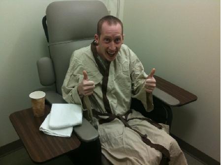
Jedi Knight Cancer Patient is hoping for good results!
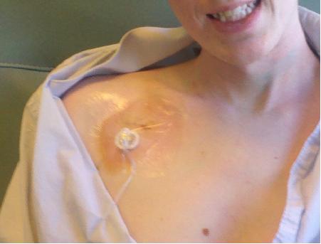
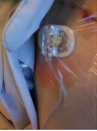
Contrast this with pictures from the last time I had a CT Scan and you can see that we’ve really come quite a ways, at least as far as my comfort level with these things goes. In fact this time the process was even easier than expected as they were able to access my chest port and use that to administer the intravenous contrast solution, saving my precious arm veins from another IV.
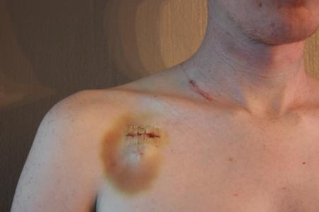
As they were “plugging me in” it occurred to me that I had yet to really talk about my chest port since the surgery last month. To start, you may want to a take moment to remind yourself what it looks like on inside, before checking out these pictures of what it looks like from the outside.
This picture was taken this evening. You can see can see the chest port under my skin. It’s little discolored as it tends to bruise a bit for a few days after being accessed. It’s not like bruise caused by trauma to the site, it’s just a small collection of blood under the skin which oozed out of the port as needle is removed from the unit. At least that’s my understanding, it could also just be bleeding from the skin cause by the needle puncture. The benefit of a chest port is that they are able to use larger thicker needles, but thicker needles leave bigger holes and the skin tends to bleed a bit more than with smaller needles.
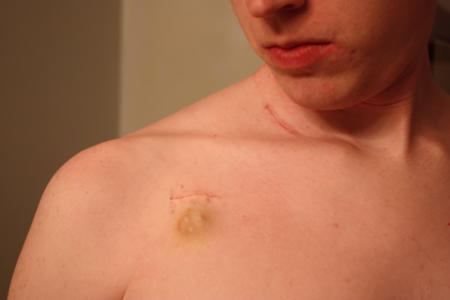
You can see the three little bumps which form a triangle, the nurses aim for the center of this triangle when they go to access the port. Directly above is the incision scar from when then put it in. Also pictured is the scar below my neck and above my collar bone from the lymph-node biopsy surgery that I had almost three months ago now. Long after I’m done with treatment I’ll have these scars to remember it by. OR, as I’ve said before, I may just come up with stories about this knife fight that I got into at a bar that one time… you should see the other guy!
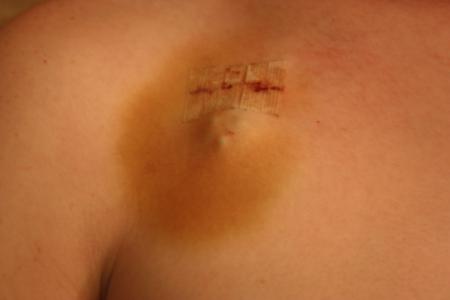
We took a few more pictures from various angles so that you can get a better idea for how it sticks out from my chest a bit.
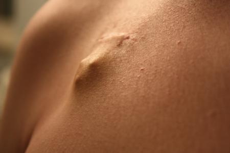
In that previous picture, the lighting is such that you can actually kind of see the catheter line a little bit too. If you create an imaginary line using the two little dots closest to the camera (so the bottom of the inverted triangle and the dot on the right), and if you follow it up the chest, you can kind of see a an ever so slight “ridge line” created by the difference in lighting on either side of it. I normally can’t see it, it’s barely visible here due to the perfect angle of the camera and the lighting. I can’t see it, but I can feel it with my fingers running up for about an inch or an inch-and-a-half where it disappears under my collar bone before running deeper into my chest.
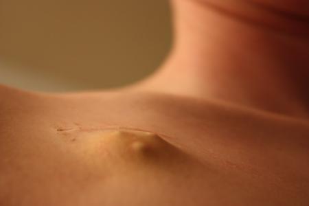
… that’s my little nubbin.
Kinda weird, isn’t it?
It’s actually really odd seeing these pictures. I’ve pretty much accepted the fact that I have this little device inside of me, but looking at these pictures… it looks like there are alien eggs, or some kind of transmitter that’s been implanted in me. At the very least you’d think I should be able to tap it like a button and ask to be “beamed up.” That’d be pretty neat. Is that too much to ask? I’d take that over a cure for cancer any day! Though I would imagine that if we did have have the early stages of Transporter technology, all of that molecular breaking down and putting back together again would probably cause cancer… but since I already have that: so bring it on!

